This example illustrates the use of the Predefined protocol for an angio-CT/SPECT fusion. It can be reproduced with data of patient P3D2 available in the example PMOD database after installation.
First the CT data was prepared. A cubic sub-volume containing the heart is extracted from the data using the Resize function available in the external tools. The remaining thoracic structures are interactively removed by drawing VOIs and setting the values outside the VOI to -1024.
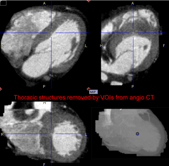
Next the SPECT data was matched to the CT in the PFUS tool, and the resliced images saved.
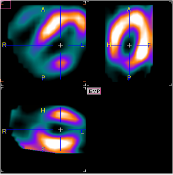
In P3D, load the prepared CT study Angio CT Reduced series of patient P3D2 and the matched SPECT. Then, select Heart (CT+ PET) from the Predefined protocols. Select Run protocol on current data and than Run Protocol button.

The protocol performs a segmentation with two RANGE thresholds (-1029-927 HU, 50-400 HU), and performs a VR rendering for each of the resulting segments.
The first segment is textured using the SPECT colors, and results in a rendering as illustrated below. To see this segment only remove the Visible check of the second and VR object activate Refresh. After adjusting the SPECT colors a bit the result looks like illustrated below
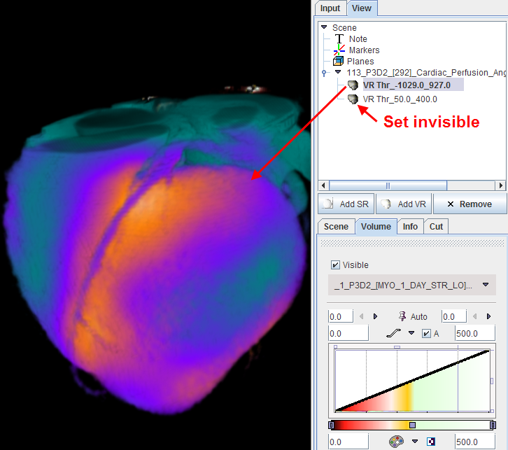
Note that the coronary arteries are colored because the SPECT study is not sharply bonded. Therefore, the second segment is used for highlighting the coronary artery structure.
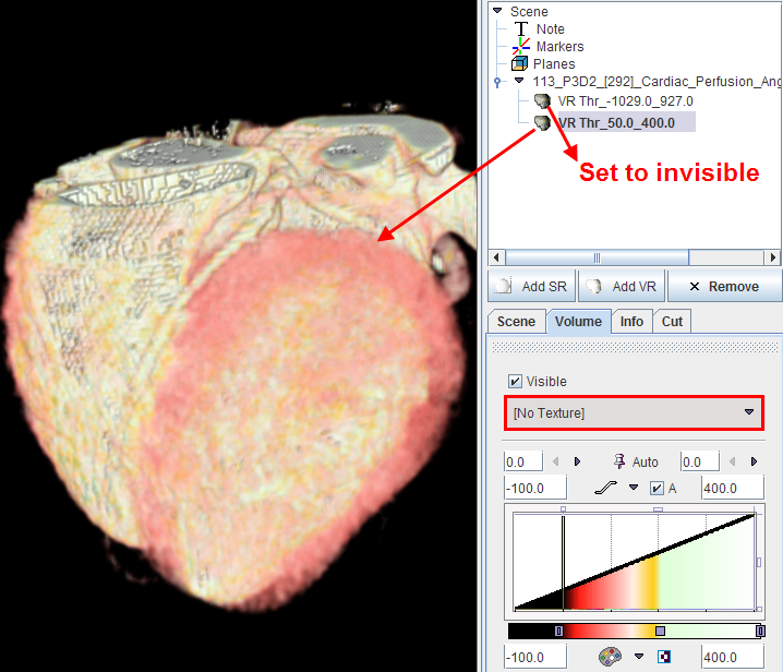
When both VR renderings are combined in a single scene (checking both Visible boxes), the following result is finally obtained.
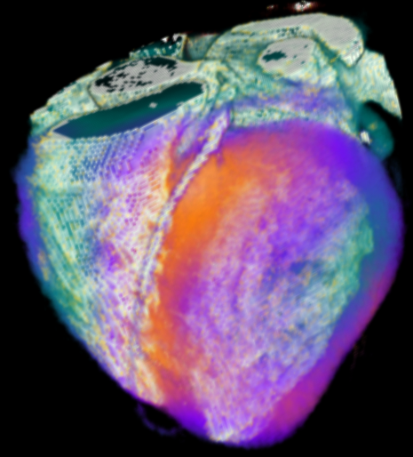
This scene could be extended by additional objects. For instance, if an important coronary artery could be individually segmented, it could be added as a clearly visible SR object.