![]()
![]()
![]()
![]()
|
|
|
|
|
Tracer in tissue is attributed to two different compartments in the following linear configuration. Tracer is taken up (K1) from arterial plasma into compartment C1. A fraction of it diffuses back to plasma (k2), another fraction moves further to compartment C2 (k3). Unless tracer is trapped in the C2 compartment (k4=0), transfer back to the intermediate compartment is also going on.
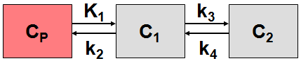
The typical interpretation is that C1 represents free and non-specifically bound tracer in tissue (non-displaceable compartment), and C2 represents specifically bound tracer.
System of differential equations:

In the auxiliary 2-tissue compartment models a direct model parameter (rate constant) is replaced by a combination of the basic parameters:
2-Tissue Compartment Model, K1/k2 |
The parameter K1/k2 (DV, distribution volume of free tracer and non-specific binding) is used as a fit parameter instead of k2, and k2 is derived from the estimated K1 and K1/k2. |
2-Tissue Compartment Model, K1/k2 & DVs |
The parameters K1/k2 and K1/k2*k3/k4 (DVs, distribution volume of specific binding) are used as fit parameters instead of k2 and k4; k2 and k4 are derived from the fit results. |
This approach allows using the non-specific distribution volume K1/k2 and DVs as common parameters in a coupled fit, as well as for the generation of synthetic curves with fixed DV and DVs.
FDG 2 Tissue Compartment Model
The FDG model is a standard 2-tissue compartment model with two additional input parameters, the lumped constant (LC) and the plasma glucose concentration (PG). In combination with the estimated K1, k2, and k3 parameters they allow to calculate the metabolic rate of glucose. When switching back and forth with the Patlak model, LC and PG are maintained, if the checkbox Model conversion in the Configuration menu is enabled.
Note: In the FDG model k4 is initially set to 0 assuming metabolic trapping, but can also be fitted.